

Secret of Light Touch: A groundbreaking study has unveiled a previously undiscovered mechanism for perceiving gentle touches: directly through our hair follicles. Until now, it was widely believed that only nerve endings in the skin and around the hair follicles were responsible for transmitting such sensations.
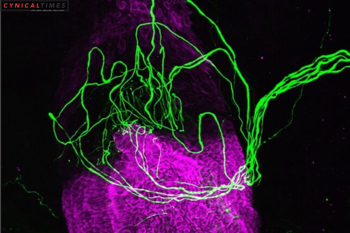
The team behind this pioneering research, led by scientists from Imperial College London in the UK, employed an advanced RNA sequencing technique. Their findings showed that cells in a part of the hair follicle known as the outer root sheath (ORS) contained a higher percentage of touch-sensitive receptors compared to equivalent cells in the skin.
Taking the investigation further, the researchers cultivated lab cultures of human hair follicle cells alongside sensory nerves. When these hair follicle cells were mechanically stimulated, the sensory nerves adjacent to them were also triggered, confirming that the sense of touch had been registered.
Also Read: Nanowire Networks Mimic Brain Learning – A Breakthrough in AI
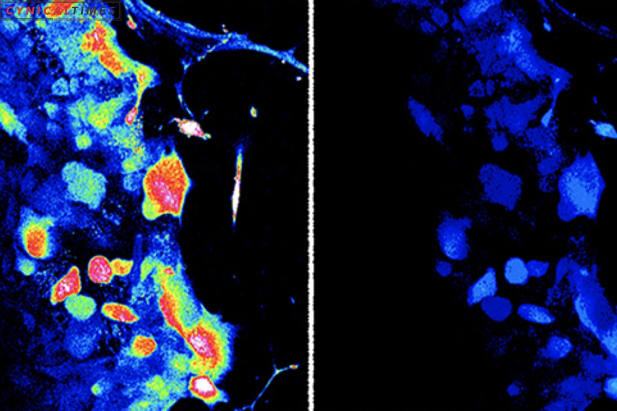
Additionally, the experiments unveiled that neurotransmitters like serotonin and histamine were released by the ORS cells through tiny vesicles, serving as signals to the surrounding cells. This discovery has opened up a realm of questions about the role of these cells and what insights they can offer into how our skin perceives touch.
Mechanoreceptors, the nerve cells responsible for touch sensation, play a crucial role in our ability to feel everything from a gentle breeze to a firm touch. In this study, the hair follicle cells interacted specifically with low-threshold mechanoreceptors (LTMRs), which are adept at sensing delicate touches.
While the importance of body hair in our sense of touch was previously acknowledged, this research has revealed a more intricate biological interaction between ORS cells and LTMRs beyond a simple mechanical response. Yet, the fundamental question remains: why do hair follicle cells possess this function in processing light touch?
“This discovery is surprising, as we are yet to unravel why hair follicle cells have this role in processing light touch,” says bioengineer Claire Higgins from Imperial College London. “Given the abundance of sensory nerve endings in the follicle, we now aim to investigate if the hair follicle activates specific types of sensory nerves for an unknown but distinct mechanism.”
Another noteworthy aspect of this research is the distinction observed when the experiments were conducted using skin cells instead of hair follicle cells. In the latter case, both histamine and serotonin were released. This suggests a unique function of ORS cells that sets them apart.
Considering the role of histamine in various inflammatory skin conditions, including eczema, it’s conceivable that further exploration of how hair follicles detect touch could lead to improved treatments and preventive measures.
Claire Higgins underscores the potential applications of their work, stating, “Our research unveils a new dimension in the release of histamine by skin cells, with promising implications for eczema research.”
This captivating research uncovers the mysterious world of touch perception, providing new insights into the unique role of hair follicles in this sensory process. It could pave the way for exciting developments in our understanding of touch and its applications in the field of dermatology.
Our Reader’s Queries
What is a light touch approach?
With her easygoing approach to office hiring rituals, she creates a comfortable atmosphere that puts everyone at ease. Her light touch and sense of humor make the process enjoyable and stress-free.
Why does light touch feel good?
Upon individual testing, it was discovered that the activation of neurons was solely attributed to norepinephrine. Merkel cells, located in the skin, are responsible for detecting light touch and transmitting signals to sensory neurons. These neurons then relay the signals to the brain for interpretation. The image provided by Ellen Lumpkin of Columbia University Irving Medical Center depicts the blue Merkel cells and red sensory neurons involved in this process.